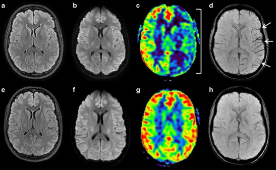
Fig. 9.

A 21-year-old patient presenting sudden-onset aphasia. Initial (top row) and control (H24, bottom row) brain MRI with FLAIR (a, e), DWI (b, f), ASL (c, g) and SWI (d, h) sequences in the axial plane. Initial MRI, a and b No visible acute ischaemic zone. c Large area of hypoperfusion affecting the whole left hemisphere (parentheses) related to migraine aura. d Left hemispheric brush sign (white arrows). Control MRI (H24) shows no ischaemic lesion (e and f), a normal left hemispheric perfusion (g), and a disappearance of the brush sign (h)
