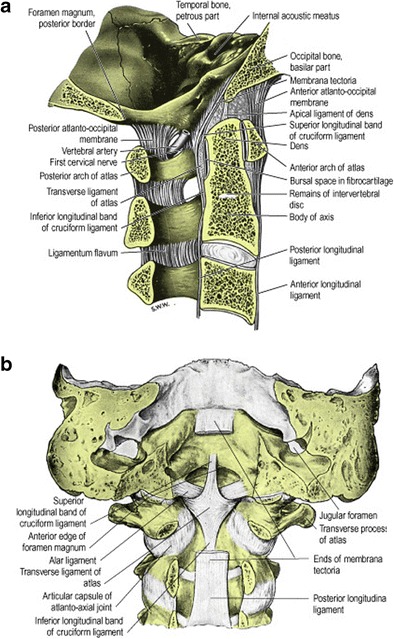
Fig. 1.

Illustrative anatomy of the craniocervical junction osseo-ligamentous structures: a right lateral view of sagittally sectioned craniocervical junction in a median plane (i.e. viewed from right to left); b posterior view of the coronally sectioned craniocervical junction; the tectorial membrane has been partly removed to expose deeper ligaments. (These illustrations were published in Gray’s Anatomy: The Anatomical Basis of Clinical Practice (39th Edition), Susan Strandring, The Back, Copyright Elsevier, 2005)
