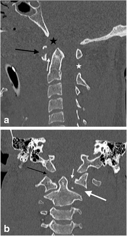
Fig. 17.

Jefferson’s fracture in a young male victim of a motor vehicle accident who suffered cardiac arrest at the scene and succumbed to his injuries shortly after presenting to the trauma centre: a sagittal reformatted CT image of the craniocervical imaging assessment demonstrates severe craniocervical junction disruption with Jefferson’s fracture (type V) of the atlas (black arrow) and associated abnormally widened predental (atlantodental); white arrow) in keeping with transverse ligament disruption and a pathologically widened basion-dens interval (black asterisk) in keeping with a distraction mechanism likely disrupting the critical alar ligaments, cruciform ligament (vertical band) and probably the tectorial membrane to some degree as well as the C1-C2 components of the ligamentum nuchae and flavum (white asterisk); b coronal reformatted CT image of the same patient confirms type II atlanto-occipital distraction (black arrow) associated with Jefferson’s fracture and disruption of the atlantoaxial joint on the left (large white arrow); the left atlantodental interval is abnormally widened (small white arrow) associated with Jefferson’s fracture of the atlas
