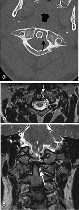
Fig. 21.

Craniocervical ligament injury in the absence of osseous injury in a 28-year-old male who sustained unimpeded fall from 3 metres associated with head injury and neck pain. There was no fracture evident on CT assessment of the craniocervical junction and sub-axial cervical spine: a axial CT image through the C1-C2 level of the craniocervical junction—there is widening of the left lateral atlanto-dental interval (black arrow) despite the absence of any fractures, raising the likelihood of craniocervical ligamentous injury; b reconstructed axial T2-SPACE MR image through the C1-C2 level of the same patient demonstrates disruption of the left transverse (atlantal) ligament (thin long white arrows) and localised haemorrhagic effusion in the widened left lateral atlanto-dental space; c coronal T2-SPACE MR image through the craniocervical junction of the same patient—the abnormal signal change related to the disrupted left transverse (atlantal) ligament and surrounding effusion is demonstrated (thick white arrow). The left alar ligament is mildly but abnormally stretched (thin white arrow). Small atlanto-axial and atlanto-occipital joint effusions are present (not annotated)
