
Fig. 3.
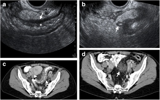
Phlegmon in a 38-year-old female with diverticulitis. a Endovaginal US reveals diffuse thickening and edema of the rectum and sigmoid colon mucosa (*) and muscular wall (arrow). b A diverticulum (arrow) with wall thickening, hyperechoic content (fecalith) and surrounding edematous fat tissue represents diverticulitis. c Axial contrast-enhanced CT of the same patient demonstrates multiple diverticulum with wall edema in distal sigmoid colon (arrow) and pericolonic fat stranding (arrowheads) representing diverticulitis. d Axial contrast-enhanced CT image at the superior level reveals a phlegmon with a hyperattenuating appearance (arrow) compared to adjacent pelvic fat