
Fig. 2.
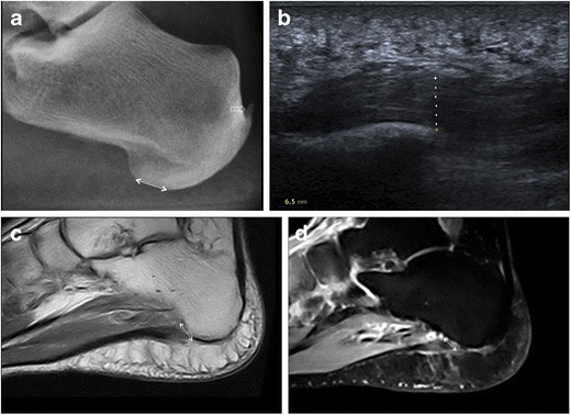
Plantar fasciitis. Lateral plain radiograph highlights an increase in the distance between subcutaneous fat and intrinsic muscles of the foot at the calcaneal insertion of the PF as an indirect sign of plantar fasciitis (double-head arrow); calcific enthesopathy of the Achilles tendon is also seen (open arrow) (a). On ultrasound, plantar fasciitis presents with PF thickening (dashed line, 6.5 mm), a hypoechoic appearance and loss of fibrillar pattern (b). MRI confirms thickening of the PF at its calcaneal origin (double-head arrow) with intrasubstance areas of intermediate and high signal intensity on T1-weighted (c) and fluid-sensitive (d) images, respectively