
Fig. 8.
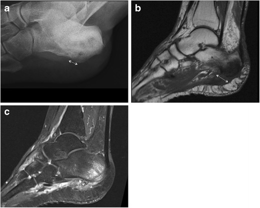
Heel osteomyelitis. Lateral plain radiograph shows marked morphological alteration of the heel with irregular lytic areas and concomitant PF thickening (double-head arrow) due to spreading of the infection (a). MRI confirms morphological alterations of the heel and PF (double-head arrow) on both T1-weighted (b) and fluid-sensitive (c) images