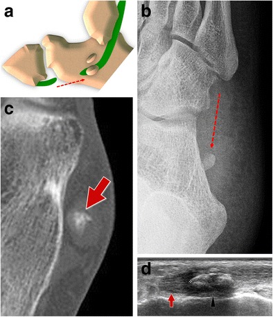
Fig. 14.

PLT tear type III standard retraction. 14A Scheme 14B Standard internal oblique radiograph. The OP shows a normal appearance but is posteriorly dislocated (generally less than 2 cm). 14C Computed tomography. CT shows a regular appearance of the displaced sesamoid bone. 14D Ultrasound. The degree of the dislocation and the size of the proximal tendinous stump (arrows) are easily measured. OP (arrowhead)
