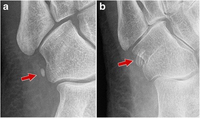
Fig. 2.

Normal os peroneum - standard radiograph. 2A Single os peroneum (arrow) visualized as an oval, well-corticated ossicle located close to the calcaneocuboid joint. 2B Bipartite os peroneum (arrow): two or more round fragments showing well-defined sclerotic margins
