
Figure 1.
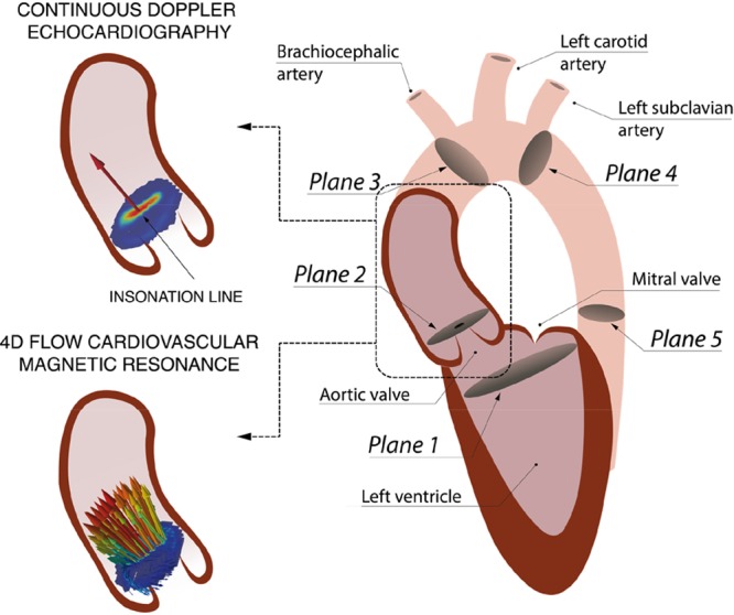
Left, Schematics of the velocity field at the vena contracta (VC) acquired during systole with continuous Doppler (1D encoded velocity value, top) and 4D flow cardiovascular magnetic resonance (CMR; 3D-encoded 2-dimensional velocity field, bottom). Right, Definition of the anatomic regions to compute the TPD from the left ventricular outflow tract (LVOT; plane 1) to the VC (plane 2). Two other anatomic regions are defined for the Material B in the Data Supplement, the ascending aorta (AA) from the VC to the brachiocephalic artery (plane 3) and the descending aorta (DA) from the left subclavian artery (plane 4) to a plane at the same height of the aortic valve plane (plane 5).