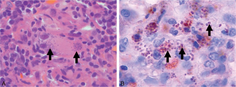
Figure 1.

Histopathology of the cervical lymphadenopathy. (A) Hematoxylin and eosin coloration (magnification ×1000) of the lymphadenopathy showing granulomatous organization with giant cells and epithelioid histiocytes. Arrows show Leishman–Donovan bodies. (B) Immunohistochemistry of the lymphadenopathy using a monoclonal anti-Leishman antibody (magnification ×1200), demonstrating leishmanial parasites with their nuclei (arrows).
