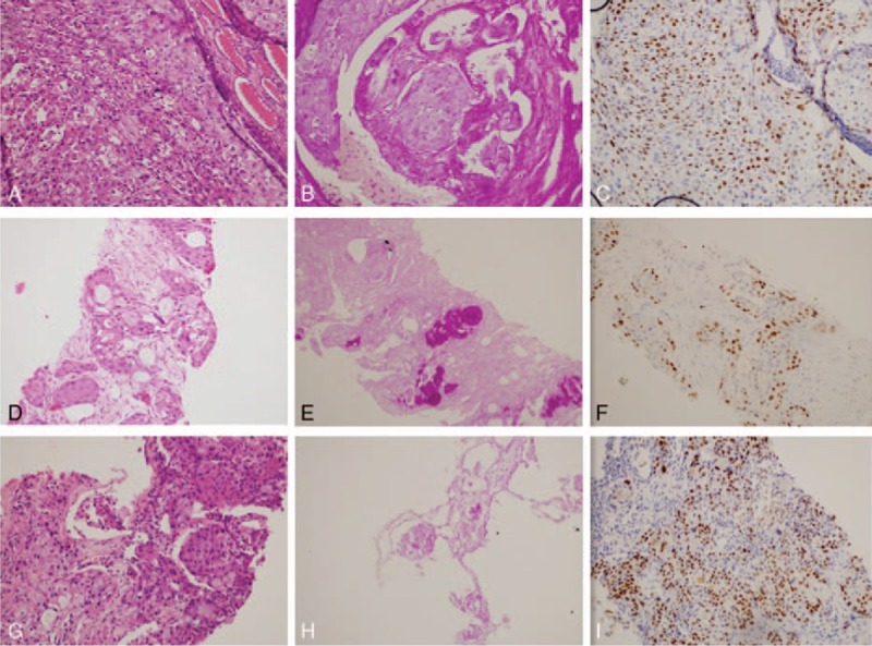
Figure 1.

Immunohistochemical staining results of thyroid, skin, and lung tumor samples. (A–C) The thyroid tumor cells in HEX200, which were positive for P63 and PAS; (D–F) immunohistochemical testing of the maculopapular eruption of the left iliac waist skin revealed the same immunohistochemical profile as that of the thyroid lesion; (G–I) the immunohistochemical testing of the lung tumor also revealed the same immunohistochemical profile. PAS, periodic acid-Schiff.
