
Figure 1.
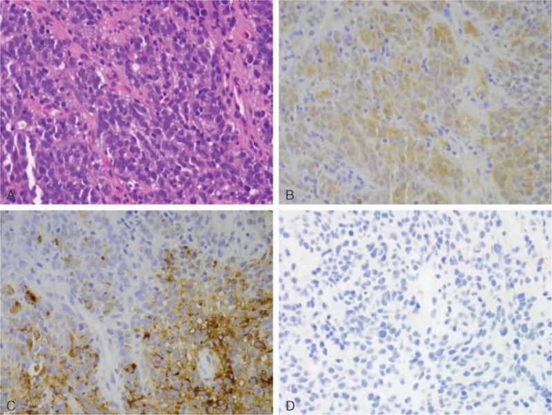
Pathohistological examination of the biopsy sample. (A) Proliferation of atypical oval to rounded cells that have hyperchromatic nuclei. Mitotic figures are frequently seen. Hematoxylin and eosin staining; magnification, ×400. (B, C, D) Immunohistochemically, the atypical tumors cells were positive for S-100 (B), HMB-45 (C) and AE1/AE3 (D). S-100, HMB-45, and AE1/AE3 staining; magnification, ×400.