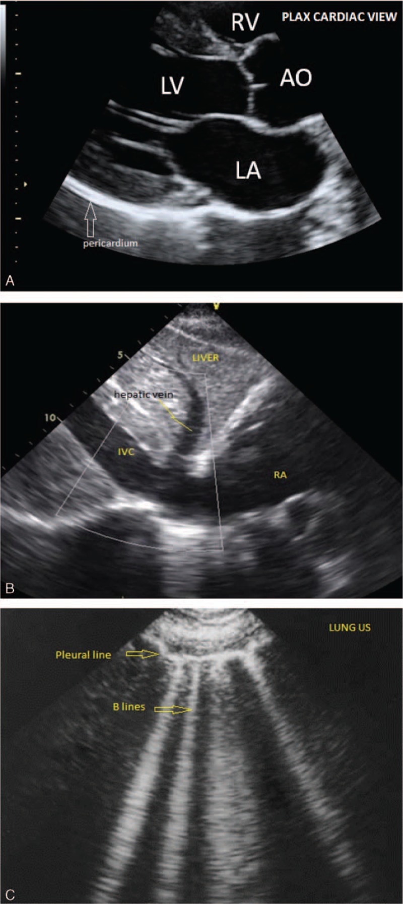
Figure 1.

Images from hand-carried ultrasound obtained during point-of-care bedside use. (A) Cardiac parasternal long axis view demonstrating right ventricle (RV), left atrium (LA), left ventricle (LV) and aorta (AO). (B) Subxiphoid view demonstrating liver, hepatic vein, inferior vena cava (IVC), and right atrium (RA). (C) Lung view demonstrating pleura and “B” lines reflecting pulmonary edema.
