Figure 1.

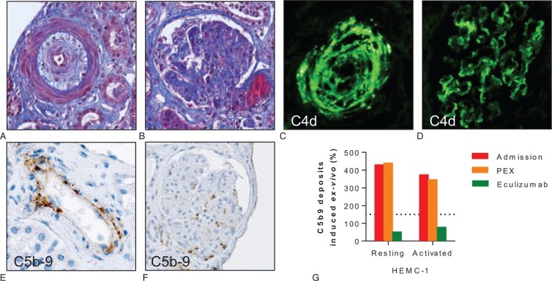
Scleroderma renal crisis and activation of the complement system. (A) Masson trichrome blue of a renal arteriole with intimal edema and onion-skin lesion narrowing the lumen (obj. ×25). (B) Masson trichrome blue of a glomerulus showing arteriolar thrombosis at the vascular pole. On light microscopy, most glomeruli appeared ischemic or even necrotic; in the remaining intact glomeruli, focal signs of mesangiolysis were present (obj. ×20). (C, D) C4d deposits identified by immunofluorescence in the wall of a renal arteriole (C) and glomerular capillaries (D). (E, F) Immunohistochemistry showing C5b-9 deposits in the endothelium of a renal arteriole (E) and a nonnecrotic glomerulus (F). (G) Ex vivo C5b-9 deposition induced by the serum of the patient. Relative surface area covered by C5b-9 staining after incubation of unstimulated (resting) or ADP-activated HMEC-1 for 4 hours with serum from the patient at admission (red bars), after 7 daily sessions of PEX (orange bars), and after 2 doses of eculizumab (green bars). C5b-9 deposits were normalized under eculizumab. Normal values <150% (dotted line). ADP = adenosine diphosphate, HMEC-1 = human microvascular endothelial cells-1, PEX = plasma exchange.
