
Figure 13.
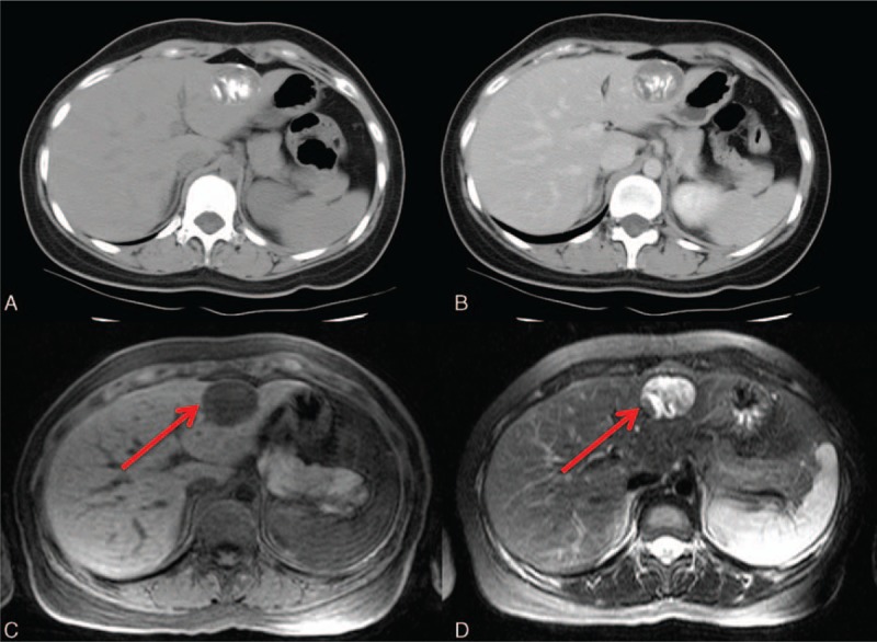
A 24-year-old woman with Echinococcus infection. A, Partially calcified hydatid cyst is well depicted on the precontrast CT image. The calcification is a consequence of the host–antigen reaction. B, The cystic component is without enhancement. C, Typical hypointensity on the T1-weighted image. D, Typical hyperintensity on the T2-weighted image. Note the characteristic hypointense rim on both the T1- and T2-weighted images (arrows in (C) and (D)), indicating the collagen produced by the host. This is the “frontline” of the host and the antigen.