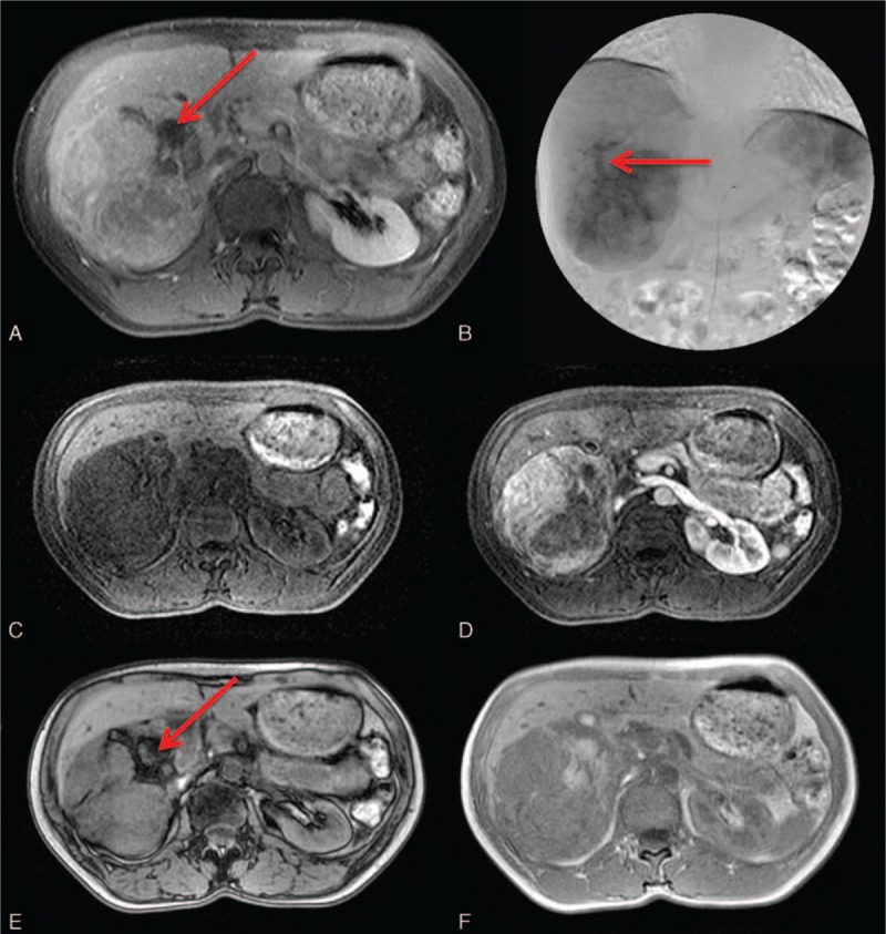
Figure 6.

A 40-year-old woman with a hepatic angiomyolipoma (AML). No significant discomfort was noted. A, The presence of macroscopic fat is clearly detected as low signal (arrow) by T1-weighted MRI with fat suppression. B, Angiography shows a draining vein (arrow) from the center of the tumor mass; this is a key characteristic of AML. Pre- (C) and post- (D) contrast T1-weighted images depict the inhomogeneous enhancement due to various degrees of angiomyomatous contents. On dual gradient-echo images, a chemical shift artifact is observed in the cancellation of the signal (arrow) on the out-of-phase (E), compared with the in-phase image (F). MRI = magnetic resonance imaging.
