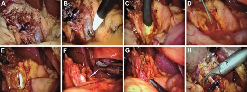
Figure 2.

(A) The cystic artery and cystic duct were dissected and ligated using absorbable clips, but for retraction purposes; the cystic duct was not divided. (B) The anterior surface of the CBD was carefully dissected for about 2.5 cm, and the CBD was performed with a longitudinal incision (8–10 mm) made with electrocautery. (C) Choledochoscope was inserted in the CBD and the left and right hepatic ducts and the distal common bile duct explored. (D) The guidewire was placed into the CBD through the choledochoscope and advanced across the papilla into the duodenum. (E) Then the guidewire was removed out until the distal end of the spontaneously removable biliary stent drainage tube entered the duodenum and the proximal end remained in the CBD. (F) The longitudinal choledochotomy was closed with 4–0 absorbable suture. (G) Bile duct suture was completed. (H) The cystic duct was divided. CBD = common bile duct.
