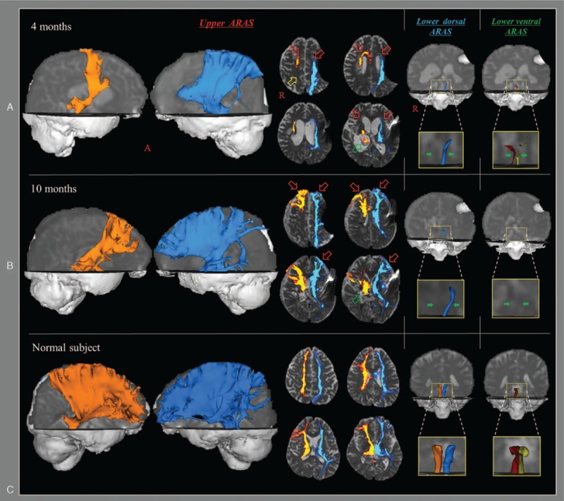
Figure 2.

Results of diffusion tensor tractography (DTT) for the ascending reticular activating system (ARAS) of the patient and a normal subject (56-year-old male). On 4-month DTT, decreased neural connectivity of the upper ARAS between the intralaminar nucleus (ILN) and the cerebral cortex was observed in both prefrontal cortexes (red arrows), right parietal cortex (yellow arrow), and right thalamus (green arrow). The lower dorsal ARAS between the pontine reticular formation (PRF) and the ILN was absent on the right side and thinner on the left side compared with the normal subject. The lower ventral ARAS between the PRF and the hypothalamus was thinner on both sides compared with the normal subject. By contrast, on 10-month DTT, marked increased neural connectivity of the upper ARAS was observed in both prefrontal cortexes (red arrows) and right thalamus (green arrows) compared with 6-month DTT. However, no significant change was observed in the lower dorsal ARAS (green arrows), and the reconstructed lower ventral ARAS (green arrows) had disappeared in both hemispheres on 10-month DTT. PRF = pontine reticular formation, ILN = intralaminar nucleus.
