
Figure 1.
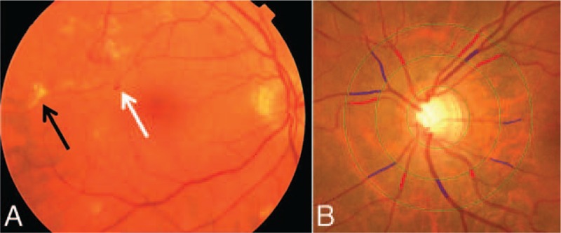
Fundus photographs showing (A) signs of retinopathy and (B) measurements of retinal vascular calibers. In (A), white arrow = small hemorrhages; black arrow = hard exudates. In (B), red lines = arteriolar calibers; blue lines = venular calibers.