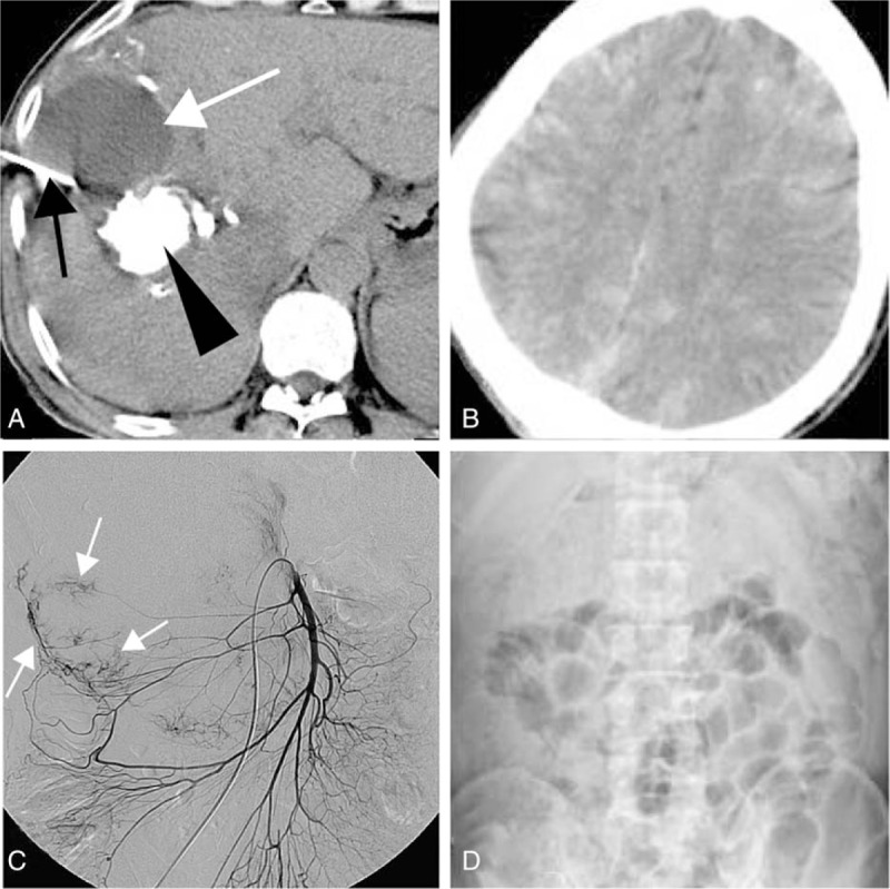
Figure 3.

(A) Computed tomography (CT) scan showed a hypodense area (white arrow). Biloma was identified by percutaneous drainage (black arrow). Lipiodol accumulated in the tumor (black arrow head); (B) noncontrast-enhanced CT scan showed multiple disseminated hyperdense lesions in the brain, consistent with deposition of lipiodol; (C and D) the superior mesenteric angiography showed tumor was fed by middle and right colic arteries (white arrow), and transarterial embolization (TACE) was performed via those arteries; partial intestinal obstruction was occurred 2 days post-TACE, and dilatation of the small bowel can be seen in the abdominal X-ray.
