. 2016 Dec 31;49(4):231–240. doi: 10.5115/acb.2016.49.4.231

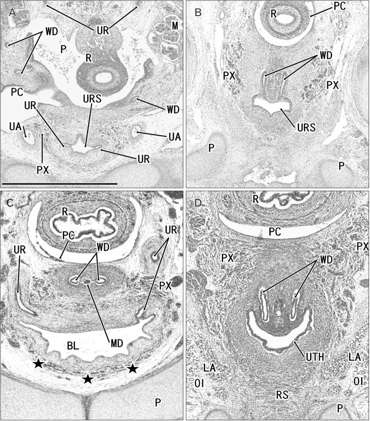
Fig. 6. Horizontal sections of two embryos at 8 weeks after differentiation of urogenital sinus into the bladder and urethra. (A, B) A specimen of crown-rump length (CRL) 29 mm. (C, D) A specimen of CRL 28 mm. Hematoxylin and eosin staining (A–C) and Orange-G staining (D). The upper side of each panel corresponds to the posterior side of the pelvis. Panels (A) and (C) display the plane 0.3 mm superior to panels (B) and (D), respectively. (C, D) In spite of the similar epithelium between sites, smooth muscles (stars) are evident in the bladder anterior wall (BL), while rhabdosphincter muscles (RS) develop along the urethra (UTH). In both specimens, the Wolffian duct (WD) opening is located 0.3 mm inferior and less than 0.1 mm medial side of the ureteral opening (UR). All panels were prepared at the same magnification. Scale bar in panel A=1 mm. BL, bladder; LA, levator ani muscle; M, mesonephros; MD, Műllerian duct or paramesonephric duct; OI, obturator internus mus c l e ; P, pubi s ; PC, peritoneal cavity; PX, pelvic autonomic nerve plexus; R, rectum; UA, umbilical artery; URS, urogenital sinus.

Fig. 6