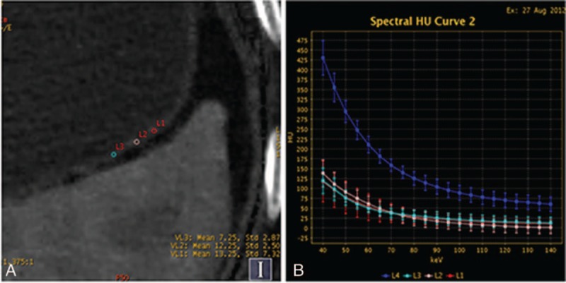
Figure 2.

Contrast enhanced images of a 60-year-old man with a right renal cyst show the iodine concentration (IC), regions of interests (ROIs), computed tomography attenuation, and spectral curve with normal gastric mucosa. (A) Iodine-based material-decomposition image at 70 keV with ROI setting on partial enlarged views during portal venous phase (PVP) and showed that IC of NGM was 13 mg/mL and normalized IC was 0.272. (B) Graph showed spectral HU curves of aorta (blue) and NGM (the other 3 colors) during PVP.
