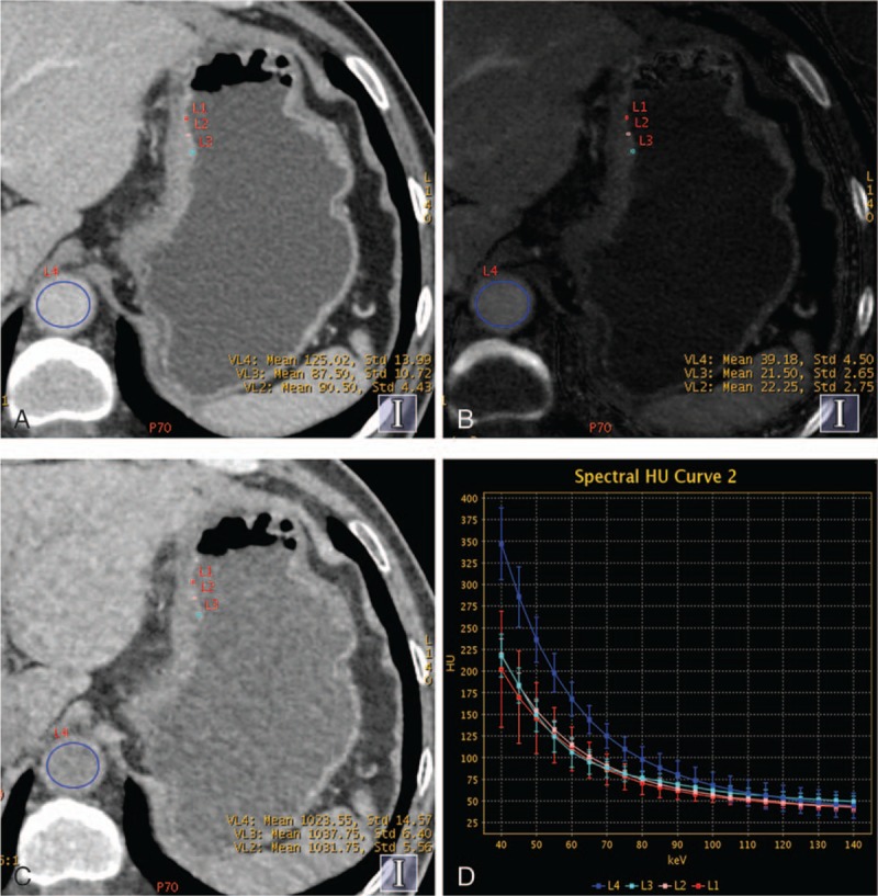
Figure 4.

Contrast enhanced gemstone spectral imaging images from a 59-year-old man with undifferentiated adenocarcinoma staged T4 in the lesser curvature of gastric in portal venous phase (PVP). (A) Monochromatic image at 70 keV during PVP showed the thickened gastric wall. (B) Iodine-based material-decomposition image showed that the iodine concentration of gastric cancer mucosa was 21.6 mg/mL, and the normalized iodine concentration was 0.55. (C) Water-based material-decomposition image. (D) Graph showed spectral HU curves of aorta (blue) and the mucosa of gastric cancer mucosa (the other 3 colors) during PVP.
