
Fig 1. Atherosclerosis in LDLR-/- and placebo DKO and caloric-restricted DKO mice.
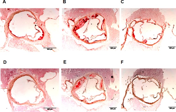
A, B, and C: Representative sections of atherosclerotic plaques are shown in which macrophages are stained with anti-MAC-3 antibody. D, E, and F: Representative sections of atherosclerotic plaques are shown in which oxidized LDL is stained with mAb4E6. Plaques in LDLR-/- mice are shown in A and D, in placebo DKO mice in B and E, and in caloric restricted DKO mice in C and F.