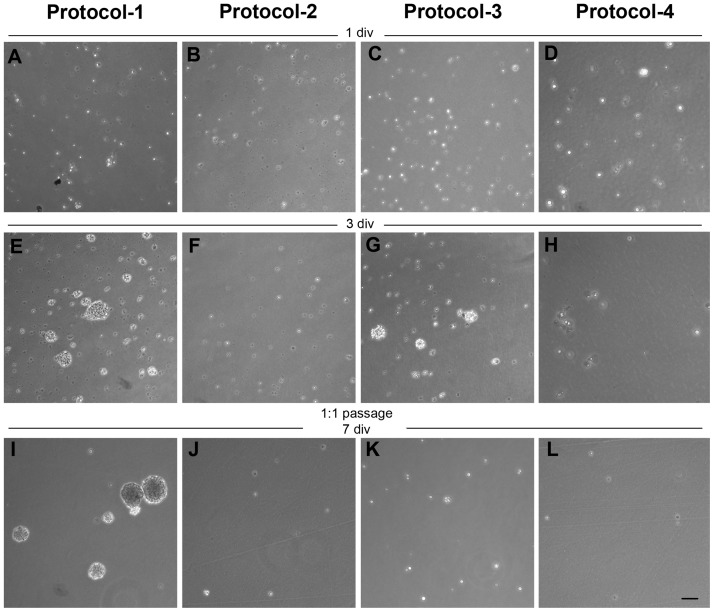
Fig 3. NS cultures of MGE-derived precursors after cryopreservation.
(A-D) Phase-contrast photomicrographs of thawed cells plated 1 day in vitro (div) in the presence of complete serum-free expansion medium. After 3 div, cells from protocol 1 (E) and 3 (G) were able to form NS. In contrast, cells from protocol 2 (F) and 4 (H) did not proliferate. Cells from protocol-1 (I) were able to form secondary NS after a 1:1 passage and 7 div, whereas cells from other protocols progressively died (J-L). Scale bar 100μm.

