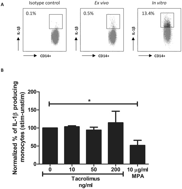
Fig 5. IL-1β production by monocytes of healthy controls is suppressed in the presence of MPA but not in the presence of tacrolimus.
(A) Dot plots showing IL-β production with or without stimulation in monocytes. Cells were gated of whole blood samples according to Fig 2a. Isotype controls were used as negative controls and were used to set the gate for the positive IL-1β expression. Results are shown as the percentage of IL-1β producing monocytes compared to the isotype control. Samples were stimulated with PMA/ionomycin for maximum production of IL-1β. (B) Mean percentages of IL-1β producing monocytes after spiking with vehicle, 10 ng/ml tacrolimus, 50 ng/ml tacrolimus or 10 μg/ml MPA. Samples were corrected for the unstimulated results and then normalized to the samples without drug exposure. IL-1β production in monocytes was significantly suppressed by a concentration of 10 μg/ml MPA. (Data are plotted as the mean ±SEM; n = 5) *) p < 0.05.

