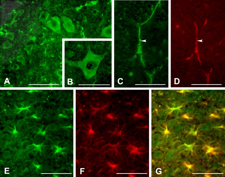
Fig 1. Immunolocalization of PAR-1 in the spinal cord of the wild-type mice.
PAR-1 is expressed by neurons in the ventral horn in the uninjured spinal cord (A). At higher magnification, the PAR-1-positive neuron exhibits typical multipolar morphology of the spinal motor neurons (B). PAR-1 (arrow, C) also co-localizes with PECAM-1-positive capillaries (arrow, D) in the uninjured cord. After spinal cord injury, PAR-1 is expressed by reactive astrocytes 24 hours post-injury (E). These reactive astrocytes in the lesion show increased expression of GFAP and hypertrophic morphology (F) as demonstrated in the digitally merged image (G). Scale bars = 100 μm for A, C, D; 50 μm for B, E, F, G.

