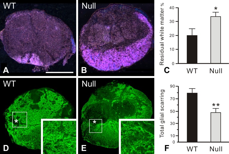
Fig 4. Enhanced white matter sparing and reduced glial scar formation at the lesion epicenter in PAR-1 null mice 42 days post injury.
Residual white matter is visualized by luxol fast blue staining using dark-field microscopy. Wild-type mice (A) have less residual white matter, mainly located at the ventral-most part of the spinal cord cross section, than the PAR-1 null mice (B). Such a difference in the size of spared white matter is statistically significant (C). The glial scar, characterized by intense GFAP immunoreactivity, is more widespread at the lesion epicenter in the wild-type mice (D) relative to the PAR-1 null mice (E). Boxed areas enclose part of the glial limitans, an interface separating the GFAP-quiescent areas (asterisks) in the lesion epicenter from the residual cord tissue. At higher magnification, more densely entangled astrocytic processes are apparent in the wild-type mice than in the PAR-1 null mice as demonstrated in the insets. As described in Materials and Methods, the quantitative analysis demonstrates that the total score of the glial scarring, which represents the severity of glial scar formation, is significantly lower in the PAR-1 null mice than in the wild-type controls (F). Scale bar = 500 μm. (n = 7 and 5/genotype for the measurement of spared white matter and glial scarring, respectively, means ± SEM, unpaired Student's t-test, *p < 0.05, **p < 0.01).

