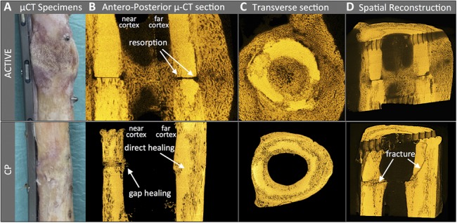
FIGURE 4.

μCT analysis after fracture due to mechanical testing: (A) Photograph of one representative specimen of each group. B, The ACTIVE specimen depicts periosteal and endosteal callus, and resorption of the osteosynthesis surface. The CP specimen depicts gap healing with transverse osteons on the plate side, and direct, primary bone healing at the far cortex. C, Transverse cross-sections adjacent to the osteotomy show cortical resorption in both specimens. D, The CP specimen illustrates a transverse fracture line through a gap-healing zone, that extends into an oblique fracture through the opposite cortex which healed by primary one healing.
