Fig. 1.
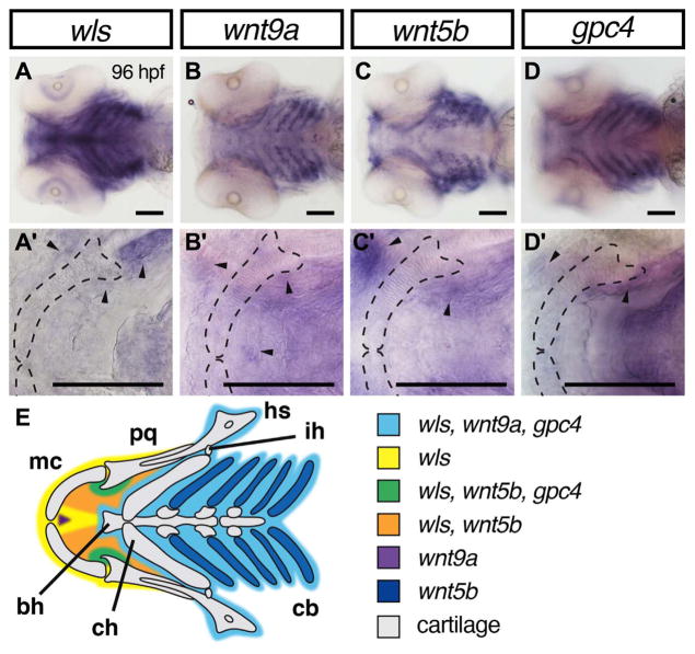
Overlapping domains of gene expression of wls, wnt9a, wnt5b and gpc4 in ventral craniofacial cartilages. (A–D) 10X Whole mount RNA in situ hybridization with ventral views with 40X focused on Meckel’s cartilage (A′–D′). At 96 hpf, wls (A) is expressed in the surrounding tissue including Meckel’s cartilage and co-expressed with wnt9a (B) and gpc4 (D) in the posterior ceratobranchials (light blue). In contrast, wnt5b appeared to be expressed in the chondrocytes of the ceratobranchials and the blood vessels surrounding them (C). wls, wnt5b and gpc4 co-localized in the surrounding mesenchyme around the jaw joint (arrowheads in A′–D′ and green in diagram E). wnt5b is expressed in the oral epithelium with wls. There is discrete expression of wnt9a in the anterior midline (purple) adjacent to the intermandibulare anterior muscle. Annotation: Meckel’s cartilage (mc), palatoquadrate (pq), hyosymplectic (hs), interhyal (ih), ceratobranchial (cb), ceratohyal (ch), basihyal (bh). Scale=50 μm.

