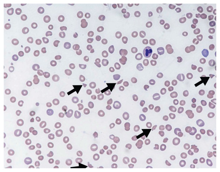
Figure 1:
Peripheral blood film showing classic findings of thrombotic microangiopathy. The image shows features of thrombocytopenia and microangiopathic hemolysis (Wright–Giemsa stain, original magnification × 50). Arrows show red blood cell fragments (i.e., schistocytes), which are substantially reduced. Image courtesy of Dr. Catherine Ross (Juravinski Hospital & Cancer Centre, McMaster University, Hamilton, Ont.).

