
FIGURE 4.
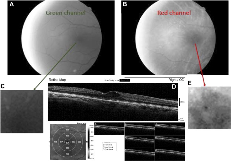
Fundus image of the right eye in a patient with ocular media changes typical of lens changes in a diabetic patient, with hard exudates in the fovea and large cysts just temporal to the foveal pit involving both the inner and outer retina. (A) Grayscale image from the green channel, with poorer contrast than the corresponding images in Fig. 1 or Fig. 3. (B) Grayscale image from the red channel of the same image, showing clearer reflectivity changes. (C) Enlarged image of the central fovea from the green-channel image. (D) OCT cross-section showing hard exudates and cysts in the fovea, and corresponding retinal thickness map. The photoreceptor layers are disrupted in the fovea. (E) Enlarged image of the central fovea taken from the red-channel image, showing the hyperreflectivity and hyporeflectivity in the central region more clearly.