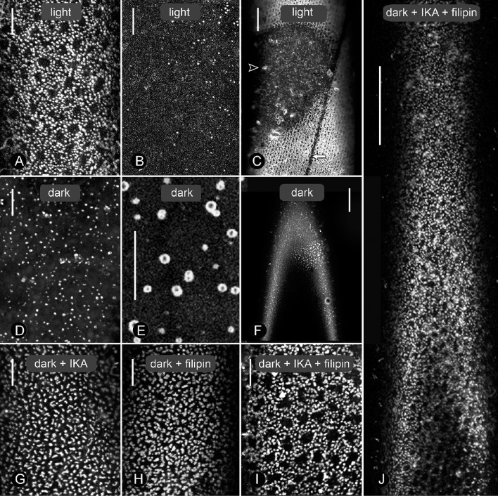
Figure 1.
Effect of dark-incubation and inhibitor treatments on size and distribution of charasomes stained by fluorescent FM1-43 in branchlet internodal cells of Chara australis. Under standard conditions of 14/10 h light/darkness, extended charasome-rich areas (A) alternate with small, charasome-poor or -free regions (B, and smaller magnification C). The white arrow in (C) indicates the neutral line, while the arrow head points to an FM-stained epiphyte. Panels (D) and (E) show different magnifications of cells exposed to continuous darkness for 12 days (control for G–J). Charasomes are mostly small and uniformly distributed (D), but “islands” with larger charasomes occasionally remained (F). (G–J) Effect of 1 μM ikarugamycin (IKA; G), 1.5 μM filipin (H), and of a combination of both inhibitors (I, J) on dark-treated cells. Note the extended charasome-rich regions (J). Bars are 10 μm (A,B,D,E,G–I) and 50 μm (C,F,J).

