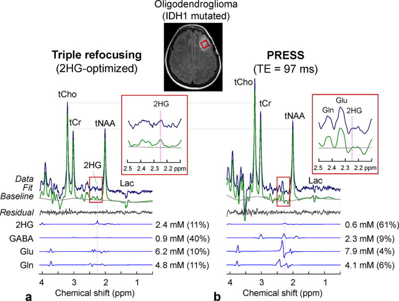
Figure 7.

In-vivo spectra from an IDH1-mutated oligodendroglioma, obtained with (a) triple refocusing and (b) PRESS TE = 97 ms (11), are shown with LCModel outputs and spectra of 2HG, GABA, Glu and Gln. The voxel size and scan time were identical between the scans (2×2×2 cm3 and 5 min). Spectra were normalized to STEAM TE = 13 ms water. Insets show magnified spectra between 2.1 and 2.5 ppm.
