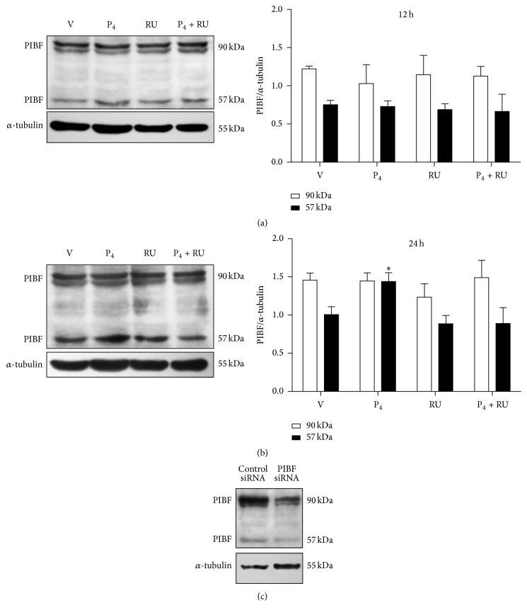
Figure 2.
PIBF (57 kDa) isoform is regulated by P4 in glioblastoma cells. Western Blot for PIBF protein was performed in U87 cells treated with vehicle (V, cyclodextrin 0.02%), P4 (10 nM), RU486 (10 μM), and P4 plus RU486 (P4 + RU) for 12 and 24 h. Representative images of PIBF isoforms content are shown with their respective densitometric analysis after 12 h (a) and 24 h (b) of treatment. For the densitometric analysis, PIBF values were corrected with those of the internal control, α-tubulin. The data are expressed as the mean ± S.E.M. with n = 4; ∗p < 0.05 versus the other treatment groups. (c) PIBF expression was silenced using a specific siRNA and a control siRNA that lacks any known mRNA target sequence. The image shows the reduction of both PIBF isoforms as evaluated by Western Blot.

