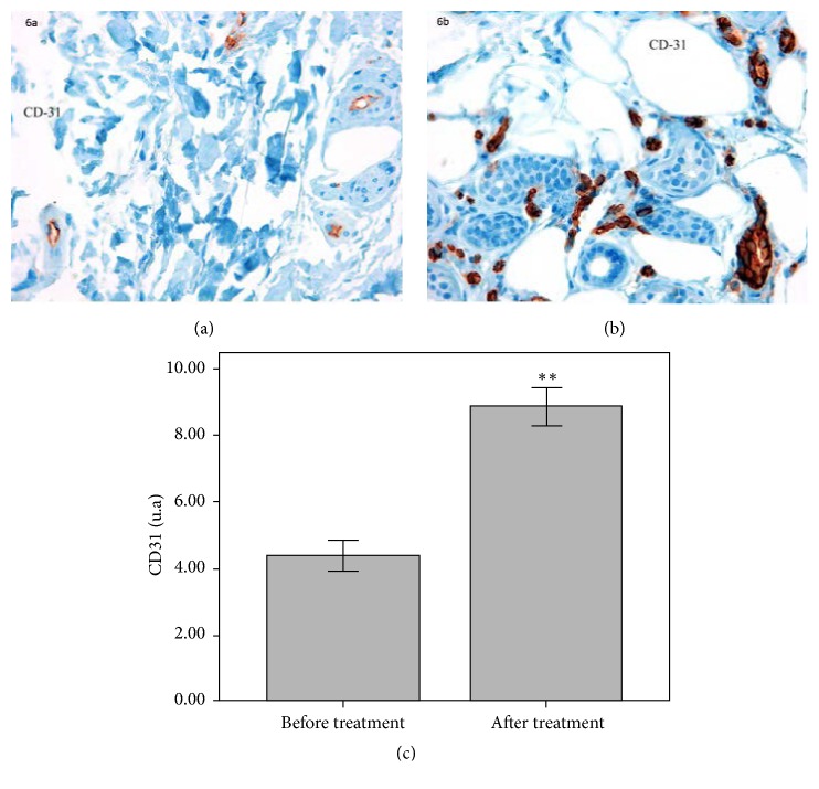
Figure 6.
Immunohistochemical staining of the endothelial marker CD-31 before (a) and after treatment (b) showing the increase in vascular presence after cosmetic treatment (40x magnification). Quantification of CD31 positive cells showed a 102.3% increase after treatment compared to baseline (∗∗p ≤ 0.01) (c).

