Figure 4.
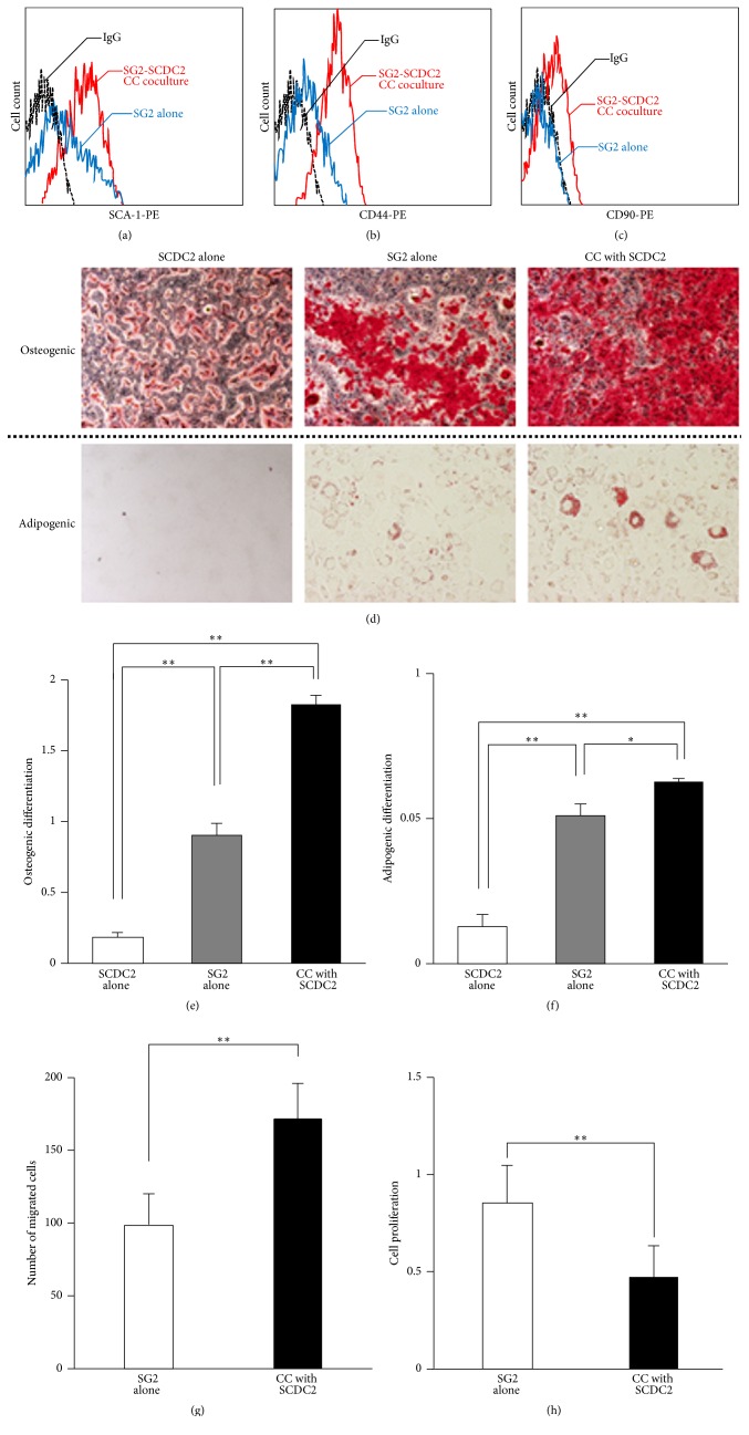
MSC stemness of SG2 cells is enhanced by direct coculture with SCDC2 cells. Cell-surface expression levels of SCA-1 (a), CD44 (b), and CD90 (c) were analyzed with each mouse-specific antibody in SG2 cells alone (blue), SG2 cells directly cocultured with SCDC2 cells (red), and an isotype control IgG (black) using flow cytometry. (d) SG2 cells were directly (CC) cocultured on the fixed feeder SCDC2 cells as described in Section 2. The SG2 cells were incubated in osteogenic (upper panel) or adipogenic (lower panel) induction medium. The cells were evaluated for extracellular matrix mineralization by alizarin red or lipid droplets by Oil Red staining. (e) Alizarin red was extracted with 10% cetylpyridinium chloride and absorbance was measured at 540 nm. (f) Oil Red O stain was extracted with DMSO and absorbance was measured at 540 nm. (g) The migratory ability of CC cocultured SG2 cells was investigated by a transwell migration assay. (h) The cell proliferation of CC cocultured SG2 cells was examined by a WST-1 assay. The results are expressed as the fold change relative to the respective control (SG2 alone). Data are presented as the mean ± standard deviation. ∗P < 0.05, ∗∗P < 0.01.

