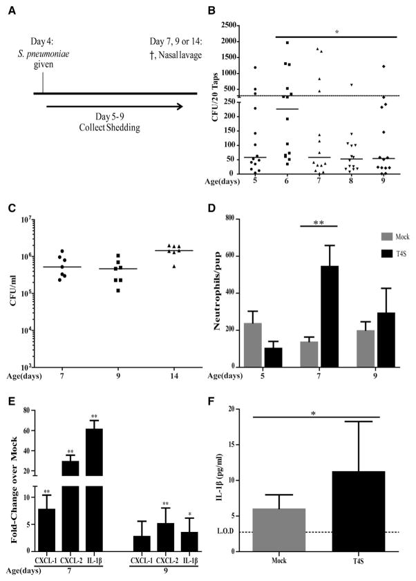
Figure 1. Pneumococcal Shedding Correlates with Upper Respiratory Tract Inflammation.
(A) 4-day-old pups were intranasally (IN) inoculated with ~2,000 CFUs S. pneumoniae strain T4S.
(B) Daily shedding was collected and quantified from nasal secretions on the days shown with median values indicated and each symbol representing the CFU observed from a single pup. Statistical analysis compares shedding on ages 6 and 9 days. Dashed line represents the 300 CFU threshold level described in the Results.
(C) Colonization density for T4S in cultures of upper respiratory tract (URT) lavages obtained from pups at the age indicated with the median value shown.
(D) Numbers of neutrophils as determined by flow cytometry (CD45+, CD11b+, and Ly6G+ events) in URT lavages obtained at the age indicated in colonized pups. Values are ±SEM (n = 5–11).
(E) The URT was lavaged with RLT RNA lysis buffer to isolate RNA from the epithelium to make cDNA from T4S colonized pups. Gene expression relative to PBS (mock)-inoculated mice was measured by qRT-PCR for the chemokine/cytokine shown. Values are ±SEM (n = 5–8).
(F) IL-1β measured by ELISA in URT lavages obtained at age 7 days for T4S or PBS (mock)-colonized pups. Values are ±SEM (n = 6–11).
*p < 0.05, **p < 0.01.

