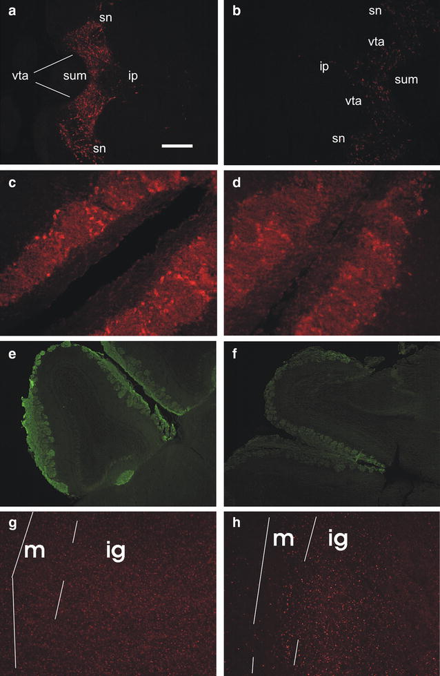
Fig. 2.

Immunohistochemistry on horizontal brain sections. Bar 200 μm for a, b, e, f; 50 μm for c, d, g, h. a–d TH immunolabelling. a Substantia nigra/ventral tegmental area appear labelled in a control mouse; caudal on the right. ip interpeduncular nucleus, sn substantia nigra, sum supramammillary nucleus, vta ventral tegmental area. b The same area is almost unlabelled in a MPTP mouse, 3 days after injections; rostral on the right. c, d Periglomerular cells are similarly labelled in a control mouse (c) and in a MPTP-treated mouse (d), 3 days after injections; caudal on top right. e, f OMP immunoreactivity in the olfactory bulb of a control (e) and a MPTP mouse (f), 3 days after injections. In the control mouse, the olfactory nerve and glomerular layer are labelled; also the accessory olfactory bulb glomerular layer is labelled. Caudal on the lower right. g, h RII immunoreactivity in the main olfactory bulb of a control (g) and a MPTP-treated mouse, 3 days after injections. Caudal on the top, lateral on the left. m mitral cell layer, ig inner granular layer
