Fig. 5.

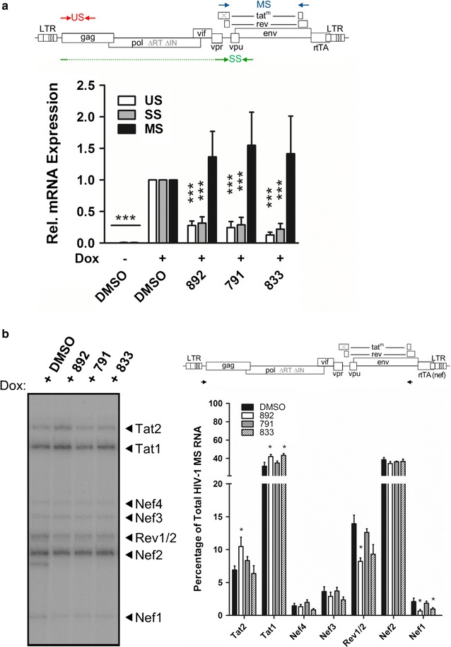
The compounds decrease the levels of HIV-1 US and SS RNAs but do not dramatically alter splice site usage. HeLa HIVrtTA∆Mls cells were incubated with 791 (30 µM), 833 (2 µM), or 892 (15 µM) for 24 h in the absence (−) or presence of (+) of Dox, then cells collected and total RNA extracted. a Top schematic of HIV-1 genome with the positions of the forward and reverse primers used for qRT-PCR analysis indicated by the arrows. US unspliced, SS singly spliced and MS multiply spliced. Bottom, quantification of viral mRNA levels in compound-treated samples were normalized to β-actin and the mean mRNA levels expressed relative to DMSO-treatment (N ≥ 7, **p ≤ 0.01, and ***p ≤ 0.001). Error bars indicate standard error of the mean (SEM). b Top, schematic of HIV-1 genome with the positions of the forward and reverse primers used to amplify the 1.8 kb class of HIV-1 RNAs indicated by the arrows. Left representative RT-PCR gel with HIV-1 MS isoforms labelled on the right according to Purcell and Martin, 1993 (N ≥ 3). Right quantification of PCR products was performed by densiometry analysis with the level of each isoform expressed as the mean percentage of the total density of all RNA species within the sample (N ≥ 7, **p ≤ 0.01, and ***p ≤ 0.001. Error bars indicate standard error of the mean (SEM)
