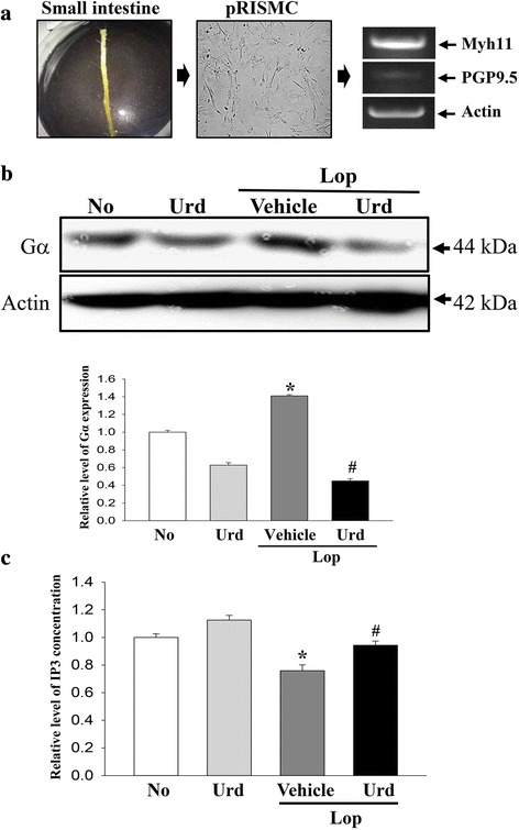
Fig. 6.

Detection of Gα expression and IP3 concentration in pRISMC. a pRISMCs were collected from the small intestines of infant rats and confirmed by RT-PCR analysis using specific primers. b Total cell lysate protein was extracted from Lop-pretreated pRISMC after treatment with the Urd. The levels of Gα expression were detected using specific antibodies. The actin level is also shown as an endogenous control. The band intensity of the three proteins was determined using an imaging densitometer and the relative level of each protein was calculated based on the intensity of actin protein as an endogenous control. b After treatment with Lop for 12 h, pRISMC were further incubated with Urd as described in the materials and methods. The IP3 concentration in the total cell lysate was measured using an ELISA kit that could detect IP3 at 5 pg/ml to 1000 pg/ml. Data represent the means ± SD of three replicates. *, p < 0.05 compared to the No treated group. #, p < 0.05 compared to the Lop + Vehicle treated group
