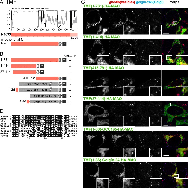
Fig. 3.

Mapping the vesicle capturing activity of TMF. a Schematic diagram of human TMF with plots for the predicted degree of coiled-coil and disorder. In the mitochondrial form, the Golgi-targeting transmembrane domain (TMD) is replaced with a hemagglutinin (HA) tag (h) and the TMD of human monoamine oxidase A (m). b Summary of the vesicle capture activity of the indicated variants of mitochondrial TMF. Capture at mitochondria was assayed by immunofluorescent staining of the Golgi integral membrane proteins golgin-84, giantin, and GalNAc-T2. Plus sign indicates that capture of all three markers was similar to the wild-type protein, minus sign indicates that no significant capture was observed. c Confocal micrographs of HeLa cells expressing the indicated TMF variants and stained for the HA tag on the golgin-84 chimera as well as for giantin that is in vesicles captured by TMF and for golgin-245, a protein that remains Golgi associated. Cells were treated with nocodazole for 6 h prior to fixation to ensure that mitochondria were close to intra-Golgi transport vesicles. Key constructs from the set shown in (b) are included, with similar results obtained using the markers golgin-84 and GalNAc-T2. Scale bars 10 μm. d Alignment of the N-terminus of human TMF with that from the indicated species. Bird, G. gallus; frog, X. tropicalis; fish D. rerio; urchin, S. purpuratus; bee, A. mellifera; oyster, C. gigas; hydra, H. vulgaris; sponge, A. queenslandica
