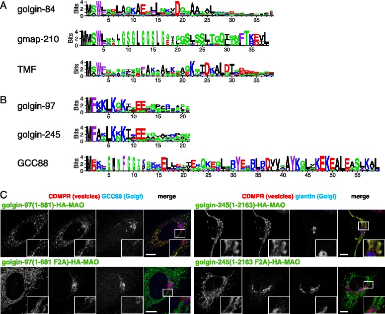
Fig. 7.

Patterns of conserved residues in the golgin vesicle capturing motifs. a Logo plots of the N-terminal regions of the three golgins that capture intra-Golgi transport vesicles. The height of the residue indicates how well it is conserved in orthologs from diverse metazoans, expressed as information content (bits). Residues are colored by their properties as follows: red, basic; blue, acidic; green, polar but uncharged; black, aliphatic; and purple, aromatic. b Logo plots of the N-terminal regions of the three GRIP domain golgins that capture endosome - to - Golgi transport vesicles. Residues as in (a). c Confocal micrographs of HeLa cells expressing the full - length mitochondrial forms of golgin-97 or golgin-245 or variants in which the conserved Phe2 residue is mutated to alanine, and stained for the hemagglutinin tag on the chimera. In both cases, this mutation results in loss of tethering of vesicles as indicated by CD-MPR. Scale bars 10 μm
