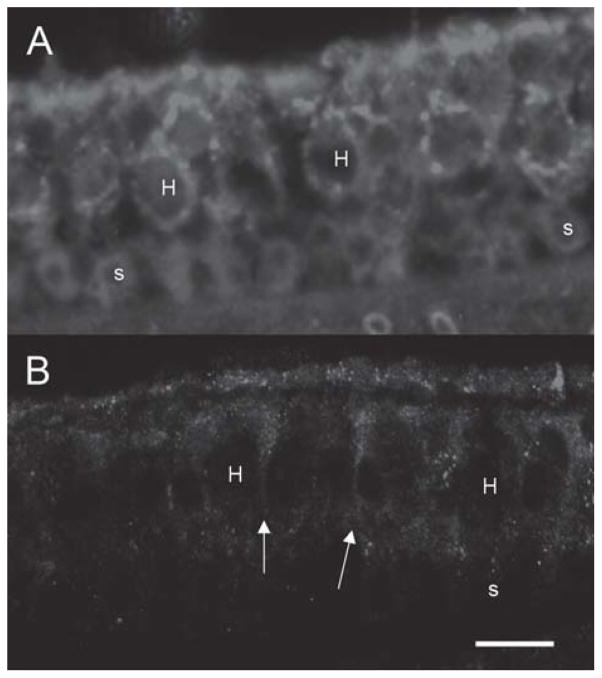
Figure 5.
Effect of a tissue-specific promoter (GFAP promoter) on GFP delivery in vivo. GFP expression using an hCMV-driven vector is seen in (A). Both hair cells and supporting cells demonstrate immunoreactivity to GFP. GFP driven by the GFAP promoter appears to weakly stain supporting cells, extending to the apical surface of the neuroepithelium (arrows). Hair cells are not stained. n=for 3 each condition. Hair cells are designated by ‘H’ and supporting cells by ‘s’. Bar = 20 μm.

