Fig. 3.
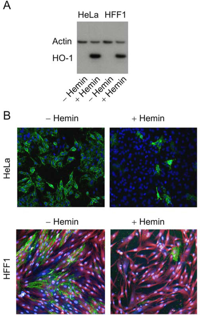
(A) Western blot analysis of hemin-treated HeLa and HFF1 cells. HO-1 expression in total-protein lysates isolated from HeLa cells and HFF1 cells cultured for 24 hours in the absence or presence of 100 µM hemin. Cells were incubated with 100 µM hemin, cellular proteins were separated on a SDS-polyacrylamide gel, transferred to PVDF nitrocellulose membrane, and probed simultaneously with HO-1 and actin antibodies. (B) Confocal microscopy of HeLa and HFF1 cells infected with EBOV under BSL-4 conditions in the absence or presence of 100 µM hemin. Images were acquired using the PE Opera confocal platform with a 10× objective. The data were analyzed using Acapella software. Green: Ebola infection; blue: nuclei (DRAQ5 stain); magenta: cytoplasm (HFF1 cells).

