. 2016 Dec 28;14(1):1–16. doi: 10.21873/cgp.20015

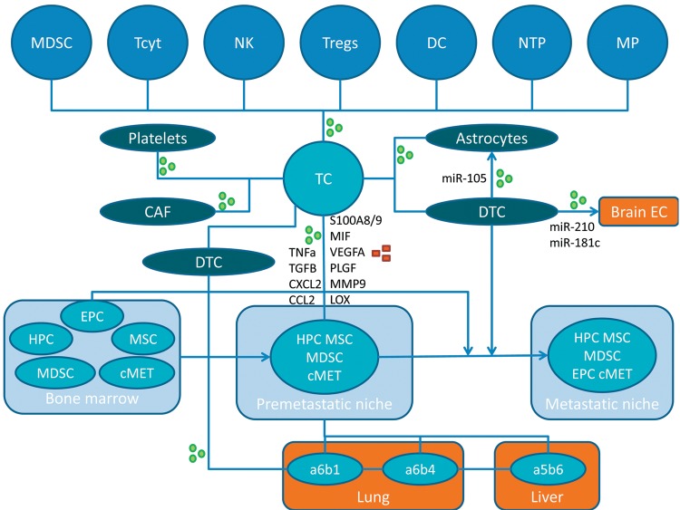
Figure 1. Interplay between tumor cells, other cell types, secreted factors and exosomes in the creation of pre- and metastatic niches. Exosomes and secreted factors are indicated by green circles or red squares, respectively. α5β6, α6β1 and α6β4,integrins; CAF, cancer-associated fibroblast; CCL2, CC chemokine ligand 2; cMET, tyrosine kinase receptor c-MET; CXCL2, chemokine (C-X-C motif) ligand 2; DC, dendritic cell; DTC, disseminated tumor cell; EC, endothelial cell; EPC, endothelial progenitor cell; HPC, hematopoietic progenitor cell; LOX, lysyl oxidase; MDSC, myeloid-derived suppressor cell; MIF, macrophage migration inhibitory factor; MMP9, matrix metalloprotease 9; MP, macrophage; MSC, mesenchymal stem cell; NK, natural killer cell; NTP, neutrophile; PLGF, pacental growth factor; S100A8/9, S100 calcium binding protein A8 or A9; TC, tumor cell; Tcyt, cytotoxic T-cell; TGFβ, transforming growth factor β; TNFα, tumor necrosis factor α; Tregs, regulatory T-cells; VEGFA, vascular endothelial growth factor, isoform A.

Figure 1