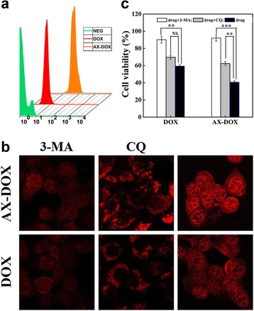
Fig. 4.

In vitro evaluation of the cellular uptake of micelles. a In vitro cellular uptake tested by flow cytometry for different DOX formulations. b CLSM images of MCF-7 cells. The cells were treated with AX-DOX, DOX at a concentration of 2 μg/mL and co-incubated with 10 mM 3-MA or 60 μM CQ for 8 h at 37 °C, respectively. c The in vitro cytotoxicity of the different DOX formulations under 10 mM 3-MA or 60 μM CQ to MCF-7 tumor cells. Data are expressed as mean ± SD (n = 3). *p < 0.05; **p < 0.01; ***p < 0.001; NS not significant
