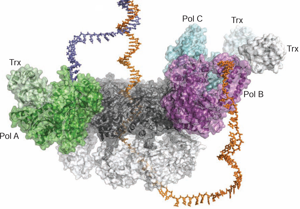
Figure 6.
Model of DNA binding in the T7 Replisome
Docking model of the T7 replisome complex bound to a replication fork DNA. The leading and lagging strands are colored slate blue and orange, respectively. As single-stranded DNA passes through the central channel of the helicase, the displaced leading strand is diverted to Pol A for leading strand synthesis. The modeled lagging strand DNA is redirected by a replication loop to interact with Pol B. In this way, the crystal structure of the T7 replisome is suggestive of a simple mechanism for coupled replication of two antiparallel DNA strands.

