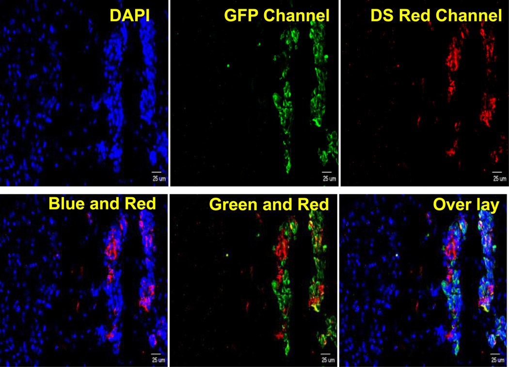
Figure 1.
Fluorescence images showing LRP Expression in MDA-MB-231Br brain metastases (Blue: DAPI to localize cell nucleus, Green: Cytokeratin to stain epithelial cells, Red: LRP 1). Primary antibodies used are Anti-Cytokeratin mouse monoclonal IgG antibody (MNF 116, Abcam) and Anti-LRP1 (low density lipoprotein receptor-related protein 1) rabbit monoclonal IgG antibody (AB 92544, Abcam). Excitation and emission filters were 470±40 and 525±50 nm for green (Alexa Fluor AF488), 560±55 and 645±75 nm for red (Alexa Fluor 594), and 320±50 and 430±60 nm for DAPI blue.

